
Atomic Images Reveal DNA Repair Enzyme
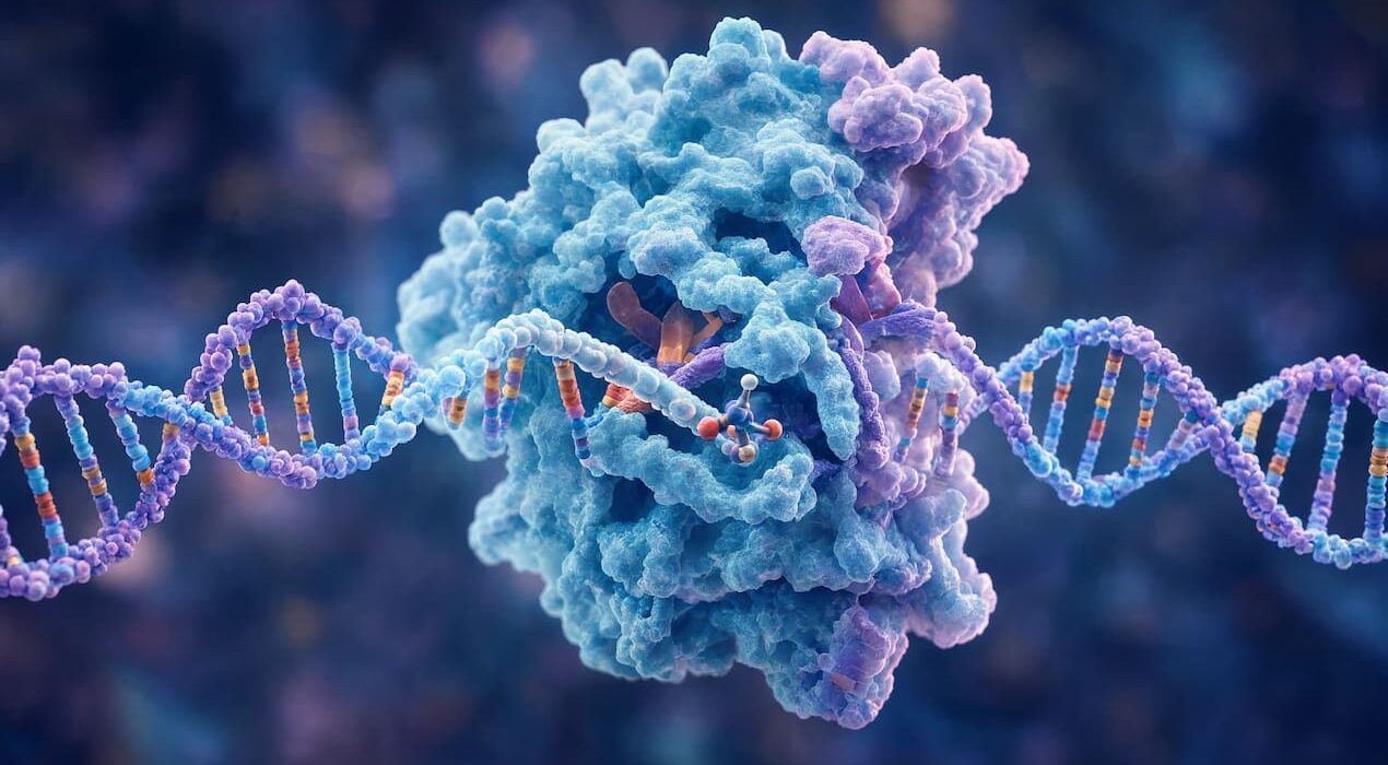
Scientists have captured the first atomic-level images of SMUG1, a human enzyme responsible for DNA repair. Every cell faces constant DNA damage from natural processes and environmental stress. If left unrepaired, these errors can cause mutations. SMUG1 acts as part of the cell’s quality control system, removing harmful bases such as uracil.
Mapping SMUG1 in Detail
Researchers at Stockholm University and partner institutes mapped SMUG1’s structure in unprecedented detail. They studied the enzyme alone, bound to uracil, attached to DNA, and interacting with chemotherapy compounds like 5‑fluorouracil. This work shows how SMUG1 identifies and removes damaged DNA components. It also reveals how the enzyme helps clear 5‑fluorouracil, a drug widely used in cancer treatment. Understanding SMUG1’s structure could guide new therapies. Drugs may one day target this repair pathway to improve cancer treatments. The study also produced the first combined neutron and X‑ray structure of a DNA‑binding protein. This rare achievement provided insight into proton positions and hydrogen‑bonding networks. Professor Stenmark explained that neutron imaging revealed details often hidden in X‑ray crystallography. The findings highlight how advanced imaging can deepen knowledge of molecular biology.
Global Collaboration
The project involved researchers from Uppsala University, Karolinska Institutet, and major European facilities such as the European Spallation Source (ESS). ESS, now under construction in Sweden, will expand opportunities for similar studies.
The Takeaway
By capturing atomic‑level images of SMUG1, scientists have revealed how cells repair DNA damage. This breakthrough offers a foundation for future therapies and strengthens our understanding of genetic stability.